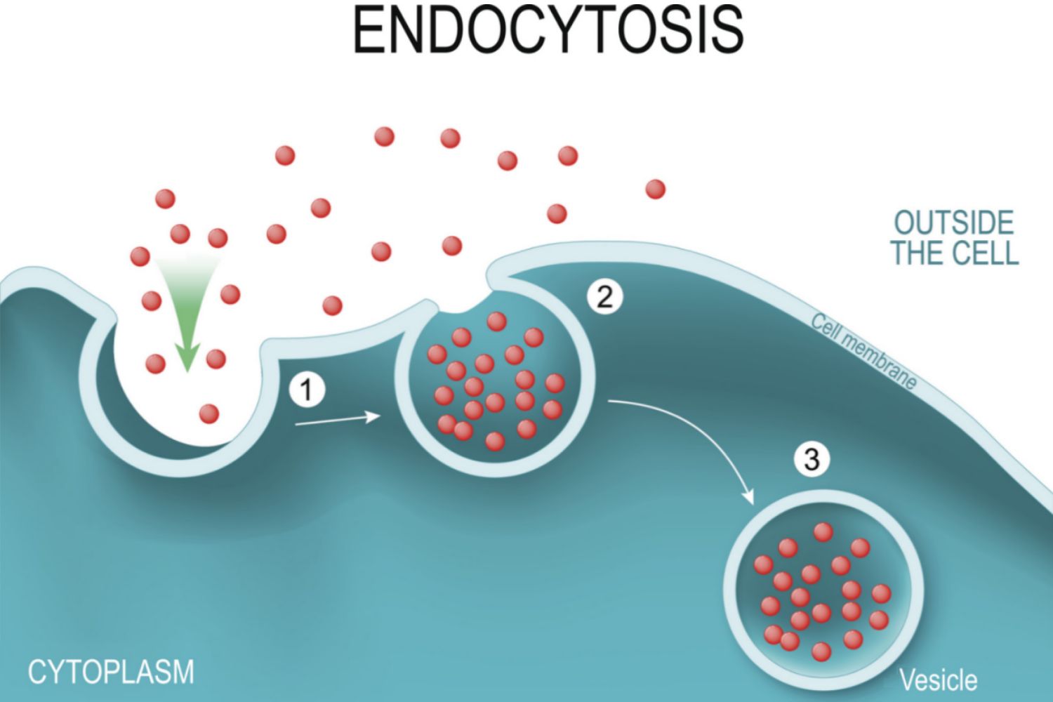
Endocytosis is the process by which cells internalize substances from their external environment. It is how cells get the nutrients they need to grow and develop. Substances internalized by endocytosis include fluids, electrolytes, proteins, and other macromolecules. Endocytosis is also one of the means by which white blood cells of the immune system capture and destroy potential pathogens including bacteria and protists. The process of endocytosis can be summarized in three basic steps.
Índice temático
The Basic Steps of Endocytosis
- The plasma membrane folds inward (invaginates) forming a cavity that fills with extracellular fluid, dissolved molecules, food particles, foreign matter, pathogens, or other substances.
- The plasma membrane folds back on itself until the ends of the in-folded membrane meet. This traps the fluid inside the vesicle. In some cells, long channels also form extending from the membrane deep into the cytoplasm.
- The vesicle is pinched off from the membrane as the ends of the in-folded membrane fuse together. The internalized vesicle is then processed by the cell.
There are three primary types of endocytosis: phagocytosis, pinocytosis, and receptor-mediated endocytosis. Phagocytosis is also called "cell eating" and involves the intake of solid material or food particles. Pinocytosis, also called "cell drinking", involves the intake of molecules dissolved in fluid. Receptor-mediated endocytosis involves the intake of molecules based upon their interaction with receptors on a cell's surface.
Lectura relacionada: Cómo Funcionan las hormonas esteroides en el Cuerpo
The Cell Membrane and Endocytosis
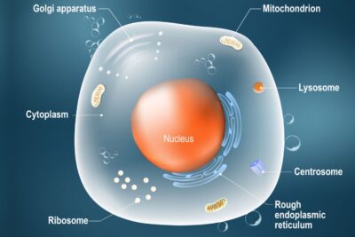
In order for endocytosis to occur, substances must be enclosed within a vesicle formed from the cell membrane, or plasma membrane. The main components of this membrane are proteins and lipids, which aid in cell membrane flexibility and molecule transport. Phospholipids are responsible for forming a double-layered barrier between the external cellular environment and the cell's interior. Phospholipids have hydrophilic (attracted to water) heads and hydrophobic (repelled by water) tails. When in contact with liquid, they spontaneously arrange so that their hydrophilic heads face the cytosol and extracellular fluid, while their hydrophobic tails move away from the fluid to the internal region of the lipid bilayer membrane.
The cell membrane is semi-permeable, meaning that only certain molecules are allowed to diffuse across the membrane. Substances that can not diffuse across the cell membrane must be helped across by passive diffusion processes (facilitated diffusion), active transport (requires energy), or by endocytosis. Endocytosis involves the removal of portions of the cell membrane for the formation of vesicles and internalization of substances. In order to maintain cell size, membrane components must be replaced. This is accomplished by the process of exocytosis. Opposite to endocytosis, exocytosis involves the formation, transportation, and fusion of internal vesicles with the cell membrane to expel substances from the cell.
Lectura relacionada: Pautas de Seguridad para Laboratorios de Biología
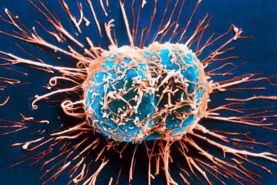
Phagocytosis
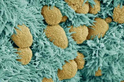
Phagocytosis is a form of endocytosis that involves the engulfing of large particles or cells. Phagocytosis allows immune cells, like macrophages, to rid the body of bacteria, cancer cells, virus-infected cells, or other harmful substances. It is also the process by which organisms such as amoebas obtain food from their environment. In phagocytosis, the phagocytic cell or phagocyte must be able to attach to the target cell, internalize it, degrade it, and expel the refuse. This process, as it occurs in immune cells, is described below.
Basic Steps of Phagocytosis
Lectura relacionada: Guía de Estudio de la Meiosis, Descripción General y Diagramas
- Detection: The phagocyte detects the antigen (substance provoking an immune response), such as a bacterium, and moves toward the target cell.
- Attachment: The phagocyte makes contact with and attaches to the bacterium. This binding initiates the formation of pseudopodia (extensions of the cell) that surround the bacterium.
- Ingestion: The surrounded bacterium is enclosed within a vesicle formed when pseudopodia membranes fuse. This vesicle with bacterium enclosed, called a phagosome, is internalized by the phagocyte.
- Fusion: The phagosome fuses with an organelle called a lysosome and becomes known as a phagolysosome. Lysosomes contain enzymes that digest organic material. The release of digestive enzymes within the phagolysosome degrades the bacterium.
- Elimination: The degraded material is expelled from the cell by exocytosis.
Phagocytosis in protists occurs similarly and more commonly as it is the means by which these organisms obtain food. Phagocytosis in humans is only performed by specialized immune cells.
Pinocytosis
While phagocytosis involves cell eating, pinocytosis involves cell drinking. Fluids and dissolved nutrients are taken into a cell by pinocytosis. The same basic steps of endocytosis are utilized in pinocytosis to internalize vesicles and to transport particles and extracellular fluid inside the cell. Once inside the cell, the vesicle may fuse with a lysosome. The digestive enzymes from the lysosome degrade the vesicle and release its contents into the cytoplasm for use by the cell. In some instances, the vesicle does not fuse with a lysosome but travels across the cell and fuses with the cell membrane on the other side of the cell. This is one means by which a cell can recycle cell membrane proteins and lipids.
Pinocytosis is nonspecific and occurs by two main processes: micropinocytosis and macropinocytosis. As the names suggest, micropinocytosis involves the formation of small vesicles (0.1 micrometers in diameter), while macropinocytosis involves the formation of larger vesicles ( 0.5 to 5 micrometers in diameter). Micropinocytosis occurs in most types of body cells and the tiny vesicles form by budding from the cell membrane. Micropinocytotic vesicles called caveolae were first discovered in blood vessel endothelium. Macropinocytosis is typically observed in white blood cells. This process differs from micropinocytosis in that the vesicles are not formed by budding but by plasma membrane ruffles. Ruffles are extended portions of the membrane that project into the extracellular fluid and then fold back on themselves. In doing so, the cell membrane scoops up the fluid, forms a vesicle, and pulls the vesicle into the cell.
Receptor-mediated Endocytosis
Receptor-mediated endocytosis is the process used by cells for the selective internalization of specific molecules. These molecules bind to specific receptors on the cell membrane before they are internalized by endocytosis. Membrane receptors are found in regions of the plasma membrane coated with the protein clatherine known as clatherine-coated pits. Once the specific molecule binds to the receptor, the pit regions are internalized and clatherine-coated vesicles are formed. After fusing with early endosomes (membrane-bound sacs that help sort internalized material), the clatherine coating is removed from the vesicles and the contents are emptied into the cell.
Basic Steps of Receptor-mediated Endocytosis
- The specified molecule binds to a receptor on the plasma membrane.
- The molecule-bound receptor migrates along the membrane to a region containing a clatherine-coated pit.
- After molecule-receptor complexes accumulate in the clatherine-coated pit, the pit region forms an invagination that is internalized by endocytosis.
- A clatherine-coated vesicle is formed, which encapsulates the ligand-receptor complex and extracellular fluid.
- The clatherine-coated vesicle fuses with an endosome in the cytoplasm and the clatherine coating is removed.
- The receptor can be enclosed in a lipid membrane and recycled back to the plasma membrane.
- If not recycled, the specified molecule remains in the endosome and the endosome fuses with a lysosome.
- Lysosomal enzymes degrade the specified molecule and deliver the desired contents to the cytoplasm.
Receptor-mediated endocytosis is thought to be more than a hundred times more efficient at taking in selective molecules than pinocytosis.
Endocytosis Key Takeaways
- During endocytosis, cells internalize substances from their external environment and get the nutrients they need to grow and develop.
- The three primary types of endocytosis are phagocytosis, pinocytosis, and receptor-mediated Endocytosis.
- In order for endocytosis to occur, substances must be enclosed within a vesicle formed from the cell (plasma) membrane.
- Phagocytosis is also known as "cell eating." It is the process used by immune cells to rid the body of harmful elements and by amoebas to obtain food.
- In pinocytosis cells "drink" fluids and dissolved nutrients in a process similar to that of phagocytosis.
- Receptor-mediated endocytosis is a much more efficient process than pinocytosis for internalizing specific molecules.
Sources
- Cooper, Geoffrey M. “Endocytosis.” The Cell: A Molecular Approach. 2nd Edition., U.S. National Library of Medicine, 1 Jan. 1970, www.ncbi.nlm.nih.gov/books/NBK9831/.
- Lim, Jet Phey, and Paul A Gleeson. “Macropinocytosis: an Endocytic Pathway for Internalising Large Gulps.” Immunology and Cell Biology, vol. 89, no. 8, 2011, pp. 836–843., doi:10.1038/icb.2011.20.
- Rosales, Carlos, and Eileen Uribe-Querol. “Phagocytosis: A Fundamental Process in Immunity.” BioMed Research International, Hindawi, 12 June 2017, www.ncbi.nlm.nih.gov/pmc/articles/PMC5485277/.
11 Tipos Diferentes de Células en el Cuerpo Humano
La Diferencia entre Mitosis y Meiosis

Biografía de Rita Levi-Montalcini, Premio Nobel

Vida de Francis Crick, Codescubridor de la Estructura del ADN
Las Células Animales y el Núcleo Unido a la Membrana
Rudolf Virchow: Padre de la Patología Moderna
Definición y Ejemplo de Células Diploides
Diferencias entre Células Vegetales y Animales
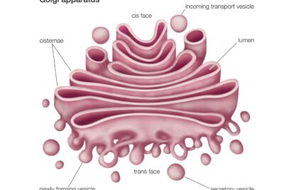
Aparato de Golgi o Complejo de Golgi

Hormonas: Definición, Tipos y Regulación
Qué Es La Difusión?
Ubicación, Estructura y Función del Plexo Coroideo