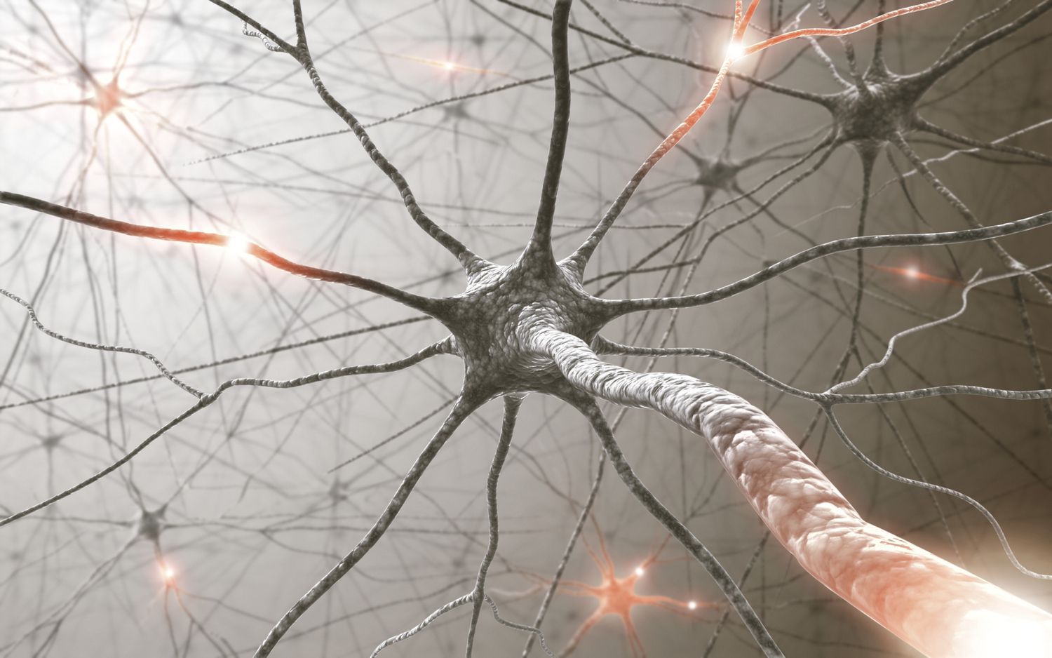
Every time you do something, from taking a step to picking up your phone, your brain transmits electrical signals to the rest of your body. These signals are called action potentials. Action potentials allow your muscles to coordinate and move with precision. They are transmitted by cells in the brain called neurons.
Key Takeaways: Action Potential
- Action potentials are visualized as rapid rises and subsequent falls in the electrical potential across a neuron’s cell membrane.
- The action potential propagates down the length of a neuron’s axon, which is responsible for transmitting information to other neurons.
- Action potentials are “all-or-nothing” events that occur when a certain potential is reached.
Action Potentials Are Conveyed by Neurons
Action potentials are transmitted by cells in the brain called neurons. Neurons are responsible for coordinating and processing information about the world that is sent in through your senses, sending commands to the muscles in your body, and relaying all the electrical signals in between.
Lectura relacionada: ¿Cómo se Relaciona la Dominancia Incompleta con el Color de Ojos?
The neuron is made up of several parts that allow it to transfer information throughout the body:
- Dendrites are branched parts of a neuron that receive information from nearby neurons.
- The cell body of the neuron contains its nucleus, which contains the cell’s hereditary information and controls the cell’s growth and reproduction.
- The axon conducts electrical signals away from the cell body, transmitting information to other neurons at its ends, or axon terminals.
You can think of the neuron like a computer, which receives input (like pressing a letter key on your keyboard) through its dendrites, then gives you an output (seeing that letter pop up on your computer screen) through its axon. In between, the information is processed so that the input results in the desired output.
Lectura relacionada:
 Las Etapas de la Mitosis y la División Celular
Definition of Action Potential
Action potentials, also called “spikes” or “impulses,” occur when the electrical potential across a cellular membrane rapidly rises, then falls, in response to an event. The entire process typically takes several milliseconds.
A cellular membrane is a double layer of proteins and lipids that surrounds a cell, protecting its contents from the outside environment and allowing only certain substances in while keeping others out.
An electrical potential, measured in Volts (V), measures the amount of electrical energy that has the potential to do work. All cells maintain an electrical potential across their cellular membranes.
Lectura relacionada: Las Leyes de la Termodinámica en los Sistemas Biológicos
The Role of Concentration Gradients in Action Potentials
The electrical potential across a cellular membrane, which is measured by comparing the potential inside of a cell to the outside, arises because there are differences in concentration, or concentration gradients, of charged particles called ions outside versus inside the cell. These concentration gradients in turn cause electrical and chemical imbalances that drive ions to even out the imbalances, with more disparate imbalances providing a greater motivator, or driving force, for the imbalances to be remedied. To do this, an ion typically moves from the high-concentration side of the membrane to the low-concentration side.
The two ions of interest for action potentials are the potassium cation (K+) and the sodium cation (Na+), which can be found inside and outside of cells.
- There is a higher concentration of K+ inside of cells relative to the outside.
- There is a higher concentration of Na+ on the outside of cells relative to the inside, about 10 times as high.
The Resting Membrane Potential
When there is no action potential in progress (i.e., the cell is “at rest”), the electrical potential of neurons is at the resting membrane potential, which is typically measured to be around -70 mV. This means that the potential of the inside of the cell is 70 mV lower than the outside. It should be noted that this refers to an equilibrium state – ions still move into and out of the cell, but in a way that keeps the resting membrane potential at a fairly constant value.
The resting membrane potential can be maintained because the cellular membrane contains proteins that form ion channels – holes that allow ions to flow into and out of cells – and sodium/potassium pumps which can pump ions in and out of the cell.
Ion channels are not always open; some types of channels only open up in response to specific conditions. These channels are thus called “gated” channels.
A leakage channel opens and closes at random and helps maintain the resting membrane potential of the cell. Sodium leakage channels allow Na+ to slowly move into the cell (because the concentration of Na+ is higher on the outside relative to the inside), while potassium channels allow K+ to move out of the cell (because the concentration of K+ is higher on the inside relative to the outside). However, there are many more leakage channels for potassium than there are for sodium, and so potassium moves out of the cell at a much faster rate than sodium entering the cell. Thus, there is more positive charge on the outside of the cell, causing the resting membrane potential to be negative.
A sodium/potassium pump maintains the resting membrane potential by moving sodium back out of the cell or potassium into the cell. However, this pump brings in two K+ ions for every three Na+ ions removed, maintaining the negative potential.
Voltage-gated ion channels are important for action potentials. Most of these channels remain closed when the cellular membrane is close to its resting membrane potential. However, when the potential of the cell becomes more positive (less negative), these ion channels will open.
Stages of the Action Potential
An action potential is a temporary reversal of the resting membrane potential, from negative to positive. The action potential “spike” is usually broken into several stages:
- In response to a signal (or stimulus) like a neurotransmitter binding to its receptor or pressing a key with your finger, some Na+ channels open, allowing Na+ to flow into the cell due to the concentration gradient. The membrane potential depolarizes, or becomes more positive.
- Once the membrane potential reaches a threshold value—usually around -55 mV—the action potential continues. If the potential is not reached, the action potential does not happen and the cell will go back to its resting membrane potential. This requirement of reaching a threshold is why the action potential is termed an all-or-nothing event.
- After reaching the threshold value, voltage-gated Na+ channels open, and Na+ ions flood into the cell. The membrane potential flips from negative to positive because the inside of the cell is now more positive relative to the outside.
- As the membrane potential reaches +30 mV – the peak of the action potential – voltage-gated potassium channels open, and K+ leaves the cell due to the concentration gradient. The membrane potential repolarizes, or moves back towards the negative resting membrane potential.
- The neuron becomes temporarily hyperpolarized as the K+ ions cause the membrane potential to become a little more negative than the resting potential.
- The neuron enters a refractory period, in which the sodium/potassium pump returns the neuron to its resting membrane potential.
Propagation of the Action Potential
The action potential travels down the length of the axon towards the axon terminals, which transmit the information to other neurons. The speed of propagation depends on the diameter of the axon—where a wider diameter means faster propagation—and whether or not a part of an axon is covered with myelin, a fatty substance that acts similar to the covering of a cable wire: it sheaths the axon and prevents electrical current from leaking out, allowing the action potential to occur faster.
Sources
- “12.4 The Action Potential.” Anatomy and Physiology, Pressbooks, opentextbc.ca/anatomyandphysiology/chapter/12-4-the-action-potential/.
- Charad, Ka Xiong. “Action Potentials.” HyperPhysics, hyperphysics.phy-astr.gsu.edu/hbase/Biology/actpot.html.
- Egri, Csilla, and Peter Ruben. “Action Potentials: Generation and Propagation.” ELS, John Wiley & Sons, Inc., 16 Apr. 2012, onlinelibrary.wiley.com/doi/10.1002/9780470015902.a0000278.pub2.
- “How Neurons Communicate.” Lumen - Boundless Biology, Lumen Learning, courses.lumenlearning.com/boundless-biology/chapter/how-neurons-communicate/.

